The translucent toolbar allows you to see the full image displayed on screen. The toolbar changes to full opacity (no transparency) when moving the mouse cursor over it. Toolbar translucency and orientation are customizable.
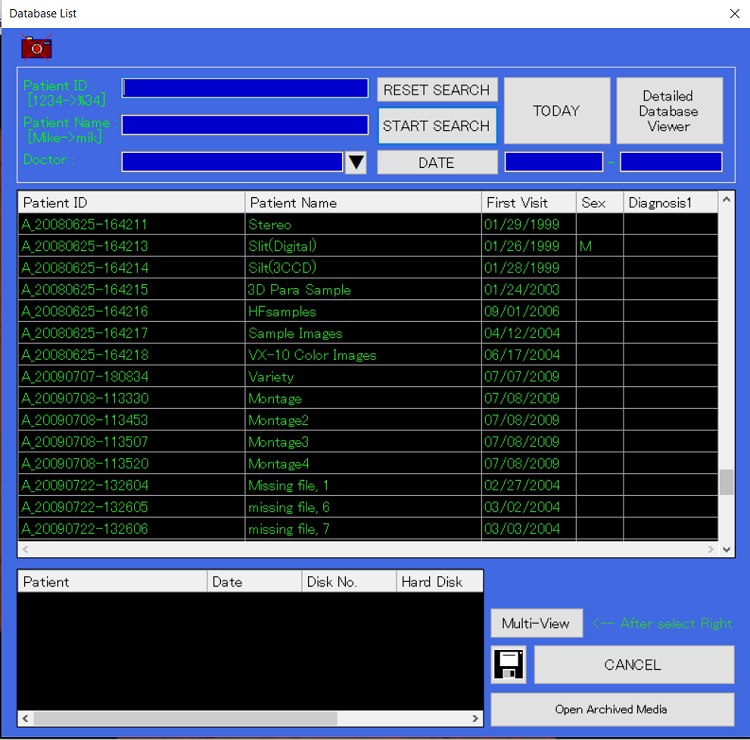
Complete display and search functions including direct input, ID list display, and pre-registration functions.
Up to 3 different digital/video devices 1-click switch.
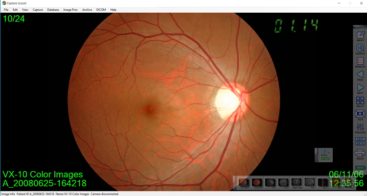
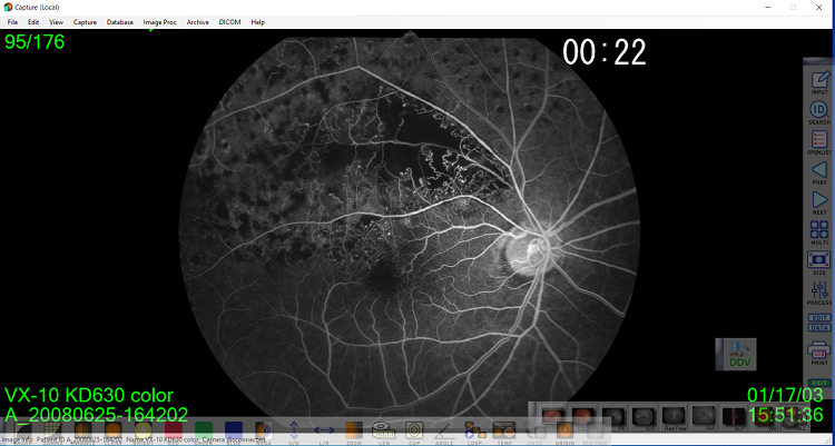
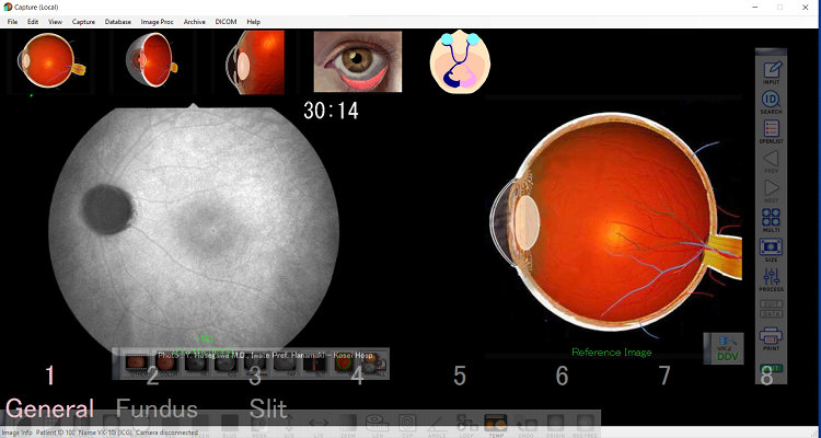
In ICG or FA mode, high speed capturing (1 frame/sec, depending on CCD camera), and the timer is displayed on the monitor (linked to Kowa retinal cameras).
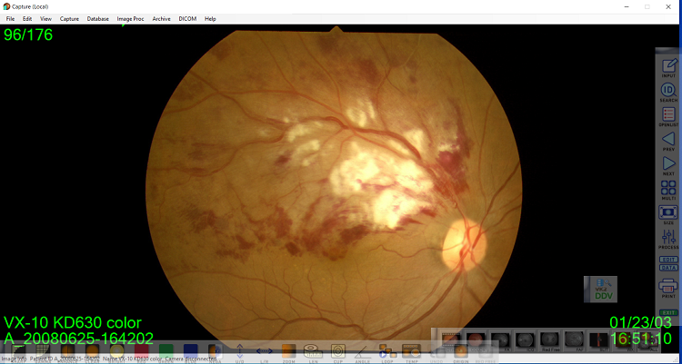
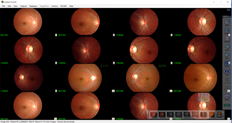
Easy analysis with 2, 4, 6, 9, 16 images displayed at the same time.
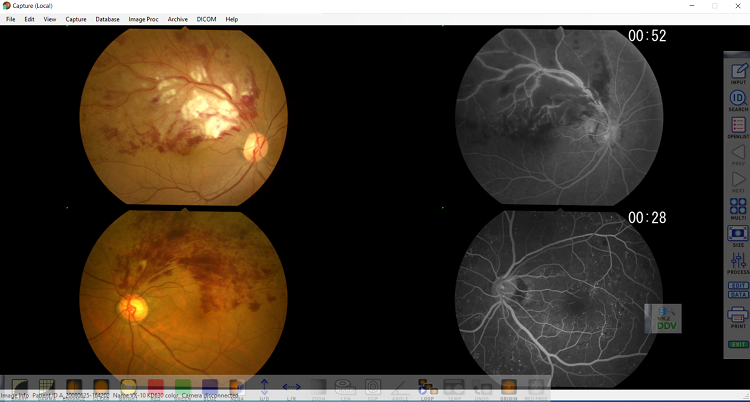
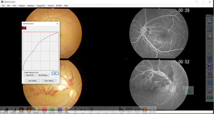
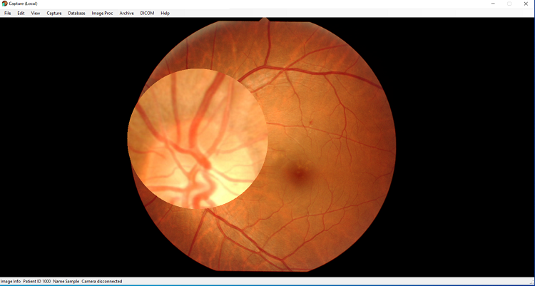
Details can be observed by left-clicking on the area to be magnified on screen.
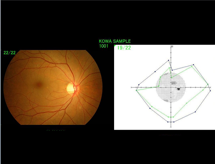
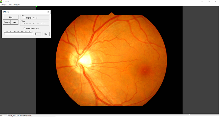
Chronological video, such as optic disc hemorrhaging, can be created from multiple still pictures for review.
View images by capture type (color, FA, ICG, slit lamp, etc.) in chronological order with a simple click on the toolbar.

Patient images can be compared with stored reference images.

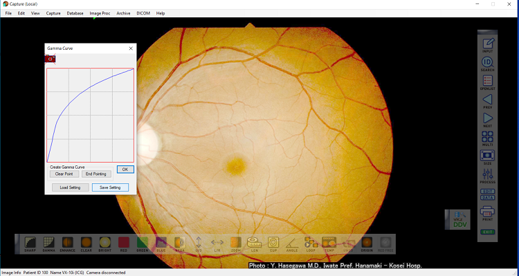
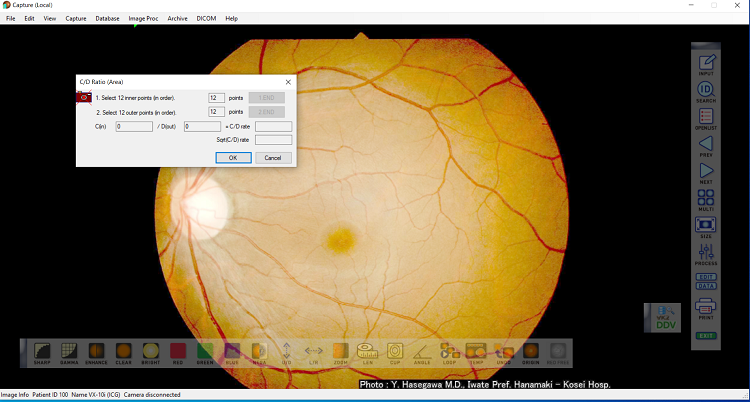
Features include: sharpen image, gamma process, enhance image, contrast, brightness, analysis with RGB filter, negative image, flip image, zoom, measure length, measure cup/disc ratio, measure angle, loop, undo, original, and reference template image.
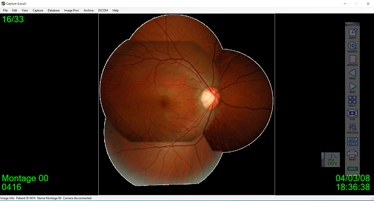
This function is enhanced with an advanced algorithm to obtain a montage of images that are easy to observe. Automatic positioning with fine rotation adjustment function is also available.
Images selected from KOWA VK-2s are easily sent to the DICOM server.
* Optional software for KOWA VK-2s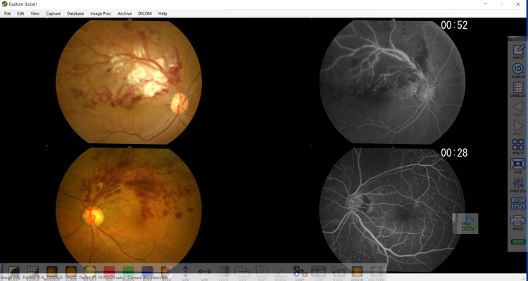
KOWA AP-7000 data can be reviewed for sharing of patient information.
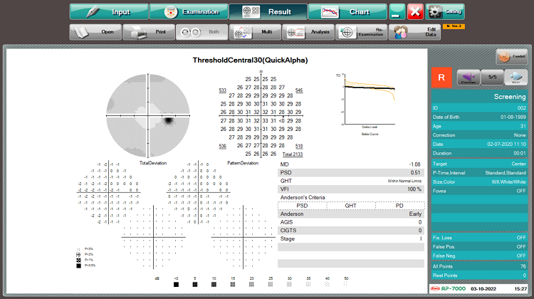
Image data captured by KOWA VK-2s can be reviewed.
